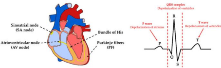
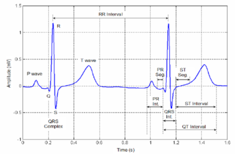
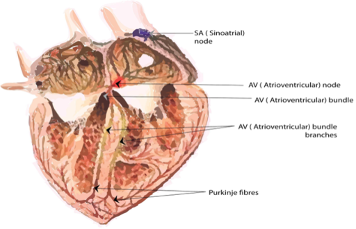
Fractal analysis is crucial for understanding complex, irregular patterns found in nature, finance, and various scientific fields. It helps to reveal self-similarity, where structures repeat at different scales, providing insights into chaotic systems like weather patterns, stock markets, and biological growth. By applying fractal analysis, researchers can model phenomena that traditional geometric methods cannot easily describe, enabling better predictions and deeper comprehension of dynamic systems. The Fractals are a fascinating mathematical tool for modeling the roughness of nature and understanding structure of such complex objects. They are considered a tool for understanding the world. In general, fractal objects are characterized by the fractal dimension. The application of fractal geometry to the analysis of ECG time series data is examined in this paper. A method based on the assessment of the Fractal Dimension (FD) of ECG recordings is suggested for the identification of cardiac diseases. In this work, and in order to exploit the fractal dimension to analyze fractal signals, the notion of fractal dimension is defined by presenting methods for calculating this dimension such as Higuchi algorithm, Katz method, regularization, box-counting etc… Each of them has its own advantages and disadvantages. This study has shown that the electrocardiogram (ECG) is a fractal signal. This allows to classify heartbeats founded on the concept of fractals. The main aim is to develop a digital technique to analyze ECG signals in order to make an accurate diagnosis of cardiovascular diseases.
| Published in | Computational Biology and Bioinformatics (Volume 12, Issue 1) |
| DOI | 10.11648/j.cbb.20241201.12 |
| Page(s) | 12-17 |
| Creative Commons |
This is an Open Access article, distributed under the terms of the Creative Commons Attribution 4.0 International License (http://creativecommons.org/licenses/by/4.0/), which permits unrestricted use, distribution and reproduction in any medium or format, provided the original work is properly cited. |
| Copyright |
Copyright © The Author(s), 2024. Published by Science Publishing Group |
Fractal Dimension, Fractal Signal, Electrocardiogram Signal, Classification of Heart Diseases, MIT/BIH Database
FD | Fractal Dimension |
ECG | Electrocardiogram |
PVC | Vickers Hardness Ventricular Complex |
PSVT | Paroxysmal Supraventricular Tachycardia |
PAC | Premature Atrial Contracture |
DS | Shrinkage According Diameter |
HS | Shrinkage According Height |
| [1] | GAO, Jianbo et XU, Bo. Complex systems, emergence, and multiscale analysis: a tutorial and brief Survey. Applied Sciences, 2021, vol. 11, no 12, p. 5736. |
| [2] | BANERJEE, Santo, EASWARAMOORTHY, D., et GOWRISANKAR, Arulprakash. Fractal functions, dimensions and signal analysis. Cham: Springer, 2021. |
| [3] | Mendis S, Puska P, Norrving B. World Health Organization. Global atlas on cardiovascular disease prevention and control. Geneva: World Health Organization (2011). |
| [4] | Kirk KJ, O’Shea J, Ruhf LK. ECG interpretation made incredibly easy!. Chris Burghardt. 5(2011). |
| [5] | Sedielmaci I, Reguig FB. Detection of some heart diseases using fractal dimension and chaos theory In 2013 8th International Workshop on Systems, Signal Processing and their Applications (WoSSPA) 1(2013): 89-94. |
| [6] | Oweis R, Hijazi L. A computer-aided ECG diagnostic tool. Computer methods and programs in biomedicine 3(2006): 279-284. |
| [7] | MIT/BIH Database available from |
| [8] | KAK, Subhash. Fractals with optimal information dimension. Circuits, Systems, and Signal Processing, 2021, p. 1-11. |
| [9] | ISLAM, Nahina, HAMID, Nafiz Imtiaz Bin, MAHMUD, Adnan, et al. Detection of some major heart diseases using fractal analysis. International Journal of Biometrics and Bioinformatics (IJBB), 2010, vol. 4, no 2, p. 63. |
| [10] | Maghsoudi F, Kiani K. A powerful novel method for ECG signal de-noising using different thresholding and Dual Tree Complex Wavelet Transform. the 2th Int. Knowledge-Based Engineering and Innovation (2015). |
| [11] | Kania M, Fereniec M, Maniewski R. Wavelet denoising for multi-lead high resolution ECG signals. Measurement science review 4(2007): 30-33. |
| [12] | Donoho DL, Johnstone IM. Adapting to unknown smoothness via wavelet shrinkage. Journal of the american statistical association 1(1995): 1200-1224. |
| [13] | Raj VN, Venkateswarlu T. ECG signal denoising using undecimated wavelet transform. In 2011 3rd International Conference on Electronics Computer Technology 1(2011): 94-98. |
| [14] | ABAJADDI, Nesrine, ELFAHM, Youssef, ZULFIQAR, Ali, et al. A robust voice activity detection based on single frequency filtering approach and the fractal dimension. 2022. |
| [15] | Kiani, Kourosh, and Farzane Maghsoudi. "Classification of 7 arrhythmias from ecg using fractal dimensions." Journal of Bioinformatics and Systems Biology 2.3(2019): 53-65. |
APA Style
Sabrine, B. A., Taoufik, A. (2024). Application of Fractal Dimension for Cardiac Arrhythmias Classification. Computational Biology and Bioinformatics, 12(1), 12-17. https://doi.org/10.11648/j.cbb.20241201.12
ACS Style
Sabrine, B. A.; Taoufik, A. Application of Fractal Dimension for Cardiac Arrhythmias Classification. Comput. Biol. Bioinform. 2024, 12(1), 12-17. doi: 10.11648/j.cbb.20241201.12
AMA Style
Sabrine BA, Taoufik A. Application of Fractal Dimension for Cardiac Arrhythmias Classification. Comput Biol Bioinform. 2024;12(1):12-17. doi: 10.11648/j.cbb.20241201.12
@article{10.11648/j.cbb.20241201.12,
  author = {Ben Ali Sabrine and Aguili Taoufik},
  title = {Application of Fractal Dimension for Cardiac Arrhythmias Classification
},
  journal = {Computational Biology and Bioinformatics},
  volume = {12},
  number = {1},
  pages = {12-17},
  doi = {10.11648/j.cbb.20241201.12},
  url = {https://doi.org/10.11648/j.cbb.20241201.12},
  eprint = {https://article.sciencepublishinggroup.com/pdf/10.11648.j.cbb.20241201.12},
  abstract = {Fractal analysis is crucial for understanding complex, irregular patterns found in nature, finance, and various scientific fields. It helps to reveal self-similarity, where structures repeat at different scales, providing insights into chaotic systems like weather patterns, stock markets, and biological growth. By applying fractal analysis, researchers can model phenomena that traditional geometric methods cannot easily describe, enabling better predictions and deeper comprehension of dynamic systems. The Fractals are a fascinating mathematical tool for modeling the roughness of nature and understanding structure of such complex objects. They are considered a tool for understanding the world. In general, fractal objects are characterized by the fractal dimension. The application of fractal geometry to the analysis of ECG time series data is examined in this paper. A method based on the assessment of the Fractal Dimension (FD) of ECG recordings is suggested for the identification of cardiac diseases. In this work, and in order to exploit the fractal dimension to analyze fractal signals, the notion of fractal dimension is defined by presenting methods for calculating this dimension such as Higuchi algorithm, Katz method, regularization, box-counting etc… Each of them has its own advantages and disadvantages. This study has shown that the electrocardiogram (ECG) is a fractal signal. This allows to classify heartbeats founded on the concept of fractals. The main aim is to develop a digital technique to analyze ECG signals in order to make an accurate diagnosis of cardiovascular diseases.
},
 year = {2024}
}
TY  - JOUR
T1  - Application of Fractal Dimension for Cardiac Arrhythmias Classification

AU  - Ben Ali Sabrine
AU  - Aguili Taoufik
Y1  - 2024/09/11
PY  - 2024
N1  - https://doi.org/10.11648/j.cbb.20241201.12
DO  - 10.11648/j.cbb.20241201.12
T2  - Computational Biology and Bioinformatics
JF  - Computational Biology and Bioinformatics
JO  - Computational Biology and Bioinformatics
SP  - 12
EP  - 17
PB  - Science Publishing Group
SN  - 2330-8281
UR  - https://doi.org/10.11648/j.cbb.20241201.12
AB  - Fractal analysis is crucial for understanding complex, irregular patterns found in nature, finance, and various scientific fields. It helps to reveal self-similarity, where structures repeat at different scales, providing insights into chaotic systems like weather patterns, stock markets, and biological growth. By applying fractal analysis, researchers can model phenomena that traditional geometric methods cannot easily describe, enabling better predictions and deeper comprehension of dynamic systems. The Fractals are a fascinating mathematical tool for modeling the roughness of nature and understanding structure of such complex objects. They are considered a tool for understanding the world. In general, fractal objects are characterized by the fractal dimension. The application of fractal geometry to the analysis of ECG time series data is examined in this paper. A method based on the assessment of the Fractal Dimension (FD) of ECG recordings is suggested for the identification of cardiac diseases. In this work, and in order to exploit the fractal dimension to analyze fractal signals, the notion of fractal dimension is defined by presenting methods for calculating this dimension such as Higuchi algorithm, Katz method, regularization, box-counting etc… Each of them has its own advantages and disadvantages. This study has shown that the electrocardiogram (ECG) is a fractal signal. This allows to classify heartbeats founded on the concept of fractals. The main aim is to develop a digital technique to analyze ECG signals in order to make an accurate diagnosis of cardiovascular diseases.

VL  - 12
IS  - 1
ER  -
Communication System Laboratory Sys'Com, National Engineering School of Tunis, University Tunis El Manar, Tunis, Tunisia