Research Article | | Peer-Reviewed

Molecular Docking, Drug-Likeness, and Toxicity Profiling of Taraxacum Officinale-derived Phytochemicals Against Estrogen Receptor Alpha (ERα) in Breast Cancer

Received: 8 September 2026     Accepted: 17 September 2026     Published: 9 October 2026
Views:       Downloads:
Abstract

Public interest in the anticancer potential of dandelion (Taraxacum officinale) root has grown following reports that its extract can induce cell death in cancer cell lines, but whether individual T. officinale phytochemicals directly engage estrogen receptor alpha (ERα), the principal target in hormone receptor-positive breast cancer, has not been characterised. This study computationally evaluated eight bioactive phytochemicals reported from T. officinale root (taraxasterol, lupeol, β-sitosterol, chlorogenic acid, caffeic acid, chicoric acid, luteolin, and quercetin) as potential ERα inhibitors, benchmarked against tamoxifen. Molecular docking was performed using AutoDock Vina against the ERα ligand-binding domain (PDB: 3ERT), with docking protocol validity confirmed by redocking the native ligand and calculating the root-mean-square deviation (RMSD). Drug-likeness was assessed using SwissADME (Lipinski's Rule of Five), and pharmacokinetic and toxicity behaviour were predicted using pkCSM. Redocking reproduced the native ligand pose with an RMSD of 0.260 Å. Binding scores ranged from -4.5 (lupeol) to -8.5 kcal/mol (luteolin), versus -9.8 kcal/mol for tamoxifen. Luteolin, quercetin, chicoric acid, β-sitosterol, and chlorogenic acid showed the most favourable docking energies; luteolin and quercetin reproduced key hydrogen bonds seen for the native ligand, though none reproduced tamoxifen's Asp351 salt bridge. Chlorogenic acid, caffeic acid, luteolin, and quercetin showed zero Lipinski violations, and all eight phytochemicals were hepatotoxicity-inactive in silico, unlike tamoxifen. These findings offer a receptor-level perspective on dandelion's reported anticancer activity and identify candidates warranting experimental validation.

Published in Computational Biology and Bioinformatics (Volume 14, Issue 2)
DOI 10.11648/j.cbb.20261402.11
Page(s) 54-69
Creative Commons

This is an Open Access article, distributed under the terms of the Creative Commons Attribution 4.0 International License (http://creativecommons.org/licenses/by/4.0/), which permits unrestricted use, distribution and reproduction in any medium or format, provided the original work is properly cited.

Copyright

Copyright © The Author(s), 2026. Published by Science Publishing Group

Keywords

Taraxacum Officinale, Estrogen Receptor Alpha, Molecular Docking, Tamoxifen, ADMET, Breast Cancer

1. Introduction
Breast cancer is the most common cancer in women across the globe and the leading cause of cancer associated mortality. The latest figures from GLOBOCAN, 2022, show 2,295,686 new cases and 665,684 deaths have been reported among women globally, with breast cancer representing nearly 23.8% of all new cancer cases and 15.4% of all cancer-related deaths among women .
Disease burden does not, however, fall equally on all regions: Asia had the highest total absolute incidence, with Europe and Northern America coming second and third, respectively, whereas Africa had the highest mortality-incidence ratio, highlighting disparities in early detection and treatment access . Globally, the burden is anticipated to rise rapidly for several decades to come, with over 6 million future cases projected to occur by 2050 , again emphasizing an immediate and ongoing requirement for therapeutics that are efficacious, affordable, and compatible with a heterogeneous disease.
Breast cancer comprises a group of molecularly distinct subtypes, each with differing clinical course and response to treatment. Using gene expression profiling, breast cancer has been further placed into discrete molecularly defined subgroups (Luminal, HER2-Enriched, Basal-like and Normal-like) with different natural history and responsiveness to therapy . Such a molecular taxonomy is for the most part captured in the clinic using hormone receptor and HER2 status, leading to four broad categories of breast cancer: hormone receptor-positive /HER2-negative cancers (closely corresponding to Luminal A subtype), hormone receptor-positive/ HER2-positive (Luminal B), HER2-enriched, and triple-negative breast cancer (TNBC). Luminal subtypes, which are defined by ER and/or PR status, are by far the most common in the clinical setting, whereas TNBC, which lacks ER, PR and HER2, accounts for 10-15% of all breast cancers and has the highest risk of recurrence with no tumour specific targeted therapies available .
Among these subtypes, ER-positive breast cancer occupies a position of particular clinical importance because it represents the largest proportion of cases and is amenable to hormonal manipulation. Reviews of ER-driven breast cancer report that approximately 70% of breast tumours are estrogen receptor-positive, making ER signalling the single most exploited therapeutic vulnerability in breast oncology . Because ER-positive disease depends on estrogen-driven proliferative signalling for tumour growth and survival, pharmacological disruption of this axis, through selective estrogen receptor modulators, receptor downregulators, or aromatase inhibitors, has become the cornerstone of both adjuvant and long-term breast cancer management.
Ligand activated transcription factor of ER, ER alpha is encoded by the gene ESR1 and is known to regulate most estrogen-induced proliferation effects in the mammary epithelium. The protein includes AF (Activation Function) domains that drive breast cancer proliferation, metastasis and is also associated with the development of drug-resistance. These can also result through a non-genomic and a ligand-independent pathways in addition to classic genomic pathway . Upon ligand binding, the receptor undergoes a conformational change that regulates recruitment of coactivator proteins to target gene promoters, driving transcriptional programmes that sustain cell cycle progression and inhibit apoptosis. Beyond its canonical role in reproductive physiology, dysregulated ERα signalling has been implicated in a range of pathological conditions, including reproductive system-related disorders such as breast, ovarian, and prostate cancer, reflecting the receptor's broad influence on hormone-responsive tissues .
The centrality of ERα to luminal breast cancer biology has made it one of the most extensively validated and clinically actionable drug targets in oncology. Its ligand-binding domain possesses a well-defined hydrophobic pocket capable of accommodating structurally diverse small molecules, a feature that has enabled the rational design of antagonists that competitively displace estradiol and lock the receptor in an inactive conformation. Structural biology has been instrumental in this effort: crystallographic analysis of the human ERα ligand-binding domain bound to the active tamoxifen metabolite 4-hydroxytamoxifen showed that antagonist binding causes helix 12 to fold back over the surface region that would otherwise recruit transcriptional coactivator proteins, physically blocking this recruitment and thereby silencing receptor-driven transcription . This structure, deposited as PDB 3ERT, remains a benchmark template for structure-based drug design targeting ERα and provides the receptor conformation used in the present docking study.
Tamoxifen, a first-generation selective estrogen receptor modulator (SERM), has been the mainstay of endocrine therapy for ER-positive breast cancer for over four decades. Mechanistically, tamoxifen and its active metabolites competitively bind the ERα ligand-binding domain, blocking the transcriptional activity of the receptor by directly binding to it, rather than degrading it, distinguishing SERMs from selective estrogen receptor downregulators such as fulvestrant . Clinically, tamoxifen's importance is difficult to overstate: it has been used for four decades as endocrine therapy and has reduced breast cancer mortality by approximately 30% , cementing its role as one of the most successful targeted therapies in the history of oncology.
Despite this success, tamoxifen therapy is constrained by significant limitations. Its efficacy is undermined by both intrinsic and acquired resistance, with approximately 60% of patients with ER-positive breast cancer eventually experiencing some form of resistance to anti-estrogen therapy over the course of treatment . Mechanistically, resistance has been linked to downregulation of ERα itself, alongside protective autophagy and dysregulated cell cycle regulators, reflecting the tumour's capacity to circumvent single-pathway blockade through multiple compensatory mechanisms . Tamoxifen's tissue-selective pharmacology further compounds these challenges: while it behaves as an antagonist in breast tissue, it exhibits agonist activity in the endometrium, a property that is characterised by dose- and duration-dependent proliferative effects on uterine tissue . Thus, long-term use of tamoxifen causes several clinically important effects; higher rates of endometrial cancer and venous thromboembolic disease , as well as frequently observed hot flashes and menopausal effects, occur among women receiving this therapy. These limitations collectively justify continued investigation into alternative or complementary ERα-targeting agents with potentially improved safety and resistance profiles.
Natural products have historically been a productive source of anticancer agents and continue to offer chemical scaffolds that are structurally distinct from synthetic libraries. Phytochemicals have been central to cancer therapeutics since the earliest antineoplastic drugs were discovered, and plant-derived compounds continue to constitute a substantial fraction of clinically used oncology drugs, exemplified by taxanes, vinca alkaloids, and camptothecin derivatives . Natural compounds are valued in oncology not only for their direct cytotoxic potential but also for their roles as chemopreventive agents and as sensitisers that can overcome multidrug resistance. Importantly, since 1981, roughly a quarter of newly approved anticancer molecules have been derived from natural sources , reflecting sustained pharmaceutical interest despite the dominance of synthetic drug discovery pipelines. This track record provides a strong rationale for systematically evaluating phytochemicals from traditionally used medicinal plants against validated cancer targets such as ERα.
Taraxacum officinale (common dandelion) is one such plant with a long history of traditional use and recent scientific interest. Ethnomedicinally, dandelion has been used across diverse cultures for centuries; it has traditionally served as a diuretic, laxative, anti-inflammatory remedy, and digestive aid, with documented use in traditional Chinese medicine and European folk medicine for liver and urinary disorders . Phytochemically, dandelion is a rich reservoir of bioactive constituents: phytochemical surveys reveal that dandelion contains a wide range of bioactive compounds, including polyphenols, phytosterols, flavonoids, carotenoids, terpenes, and coumarins, several of which act synergistically and contribute to antioxidant, antimicrobial, anti-inflammatory, and anticancer effects . More than 197 compounds have been catalogued from T. officinale alone, with recent reviews expanding the genus-wide inventory considerably .
Dandelion, especially the root extract, has been increasingly noted by the public and scientific community because of its reported anticancer effects. Aqueous dandelion root extract caused selective apoptosis in colonic cancer cells. In the original research, an aqueous dandelion root extract has been shown to cause cell death in over 95 percent of colonic cancer cells in 48 hours but in healthy cells and to cause tumor growth reduction in xenograft models after oral adminstration . This finding, alongside earlier reports of selective apoptosis induction in pancreatic cancer cells with no significant cytotoxic effect on non-cancerous fibroblasts and in drug-resistant leukemia and melanoma cell lines, has contributed to considerable public interest in dandelion root as a potential anticancer agent. However, a crude extract producing selective cytotoxicity in a cell-based assay does not, on its own, establish which individual phytochemical is responsible, nor does it identify the molecular target through which any effect is mediated, and this gap in mechanistic understanding motivates the present investigation.
This distinction matters because crude plant extracts are inherently complex mixtures containing dozens to hundreds of chemically diverse constituents that may act independently, additively, or synergistically, and whose relative concentrations vary with growing conditions, extraction method, and plant part used. Investigating individual, chemically defined bioactive compounds, rather than the whole extract, is therefore substantially more informative for mechanistic drug discovery: it allows a specific molecule to be linked to a specific molecular interaction with a specific target, enables rational structure-activity reasoning, and provides a tractable starting point for lead optimisation. Reviews of Taraxacum-derived oncotherapy candidates have specifically identified taraxasterol, chlorogenic acid, chicoric acid, and related constituents as promising agents capable of inhibiting tumour cell proliferation and modulating oncogenic pathways , underscoring that the anticancer potential of dandelion root is most plausibly attributable to a subset of its phytochemical constituents rather than to the extract as an undifferentiated whole.
Given the expense, time, and ethical burden associated with in vitro and in vivo pharmacological testing, computational approaches have become an indispensable first step in modern drug discovery. Molecular docking is one of the most commonly employed structure-based technique for this, facilitating computational modeling of the most energetically favourable position, orientation and conformation of a ligand within a target macromolecule’s binding site, and thus offering an expedient approach to ranking of prospective molecules ahead of the expensive process of wet-laboratory validation . However, the reliability of any docking protocol must first be established through validation: a standard approach involves redocking the co-crystallised ligand into its native binding site and calculating the root-mean-square deviation (RMSD) between the redocked and experimentally observed poses, with an RMSD value of ≤2.0 Å widely considered acceptable evidence of a reliable docking procedure . Once validated, the docking protocol can be extended with confidence to score and rank candidate phytochemicals against the same target.
Docking alone, however, is insufficient to prioritise a viable drug candidate. Complementary in silico pharmacokinetic and toxicological profiling is equally essential, since a compound with excellent predicted binding affinity may nonetheless be unsuitable for development due to poor absorption, metabolic instability, or toxicity liabilities. Physicochemical drug-likeness filters such as Lipinski's Rule of Five remain a standard early screen, given that compounds analysed from thousands of successful oral drugs cluster within specific, well-defined property ranges , while dedicated servers such as SwissADME and pkCSM extend this assessment to pharmacokinetic behaviour, drug-likeness, and medicinal chemistry compatibility , allowing rapid, cost-effective triage of candidate molecules prior to any experimental commitment. Toxicity prediction tools complete this pipeline by flagging potential mutagenic, hepatotoxic, or cardiotoxic liabilities early, further reducing the risk of late-stage attrition. Together, docking validation, drug-likeness assessment, ADMET prediction, and toxicity screening constitute a coherent, evidence-based in silico workflow that allows researchers to make a scientifically defensible case for or against a compound's therapeutic potential before any biological assay is performed.
Despite the accumulating evidence for the anticancer activity of Taraxacum officinale extracts, and despite the identification of specific bioactive constituents implicated in this activity, a critical gap remains in the literature: the direct molecular interaction between individual, major T. officinale phytochemicals and estrogen receptor alpha (the principal therapeutic target in hormone receptor-positive breast cancer) has not been systematically characterised. Existing anticancer studies of dandelion have relied predominantly on crude extracts and phenotypic cell-based assays, which, while valuable, do not establish whether any single constituent engages ERα in a manner comparable to established SERMs such as tamoxifen. Without this receptor-level mechanistic evidence, the relevance of dandelion's reported anticancer activity to hormone receptor-positive breast cancer specifically cannot be rationally assessed.
The aim of the present study was therefore to computationally evaluate eight bioactive phytochemicals reported from Taraxacum officinale root (taraxasterol, lupeol, β-sitosterol, chlorogenic acid, caffeic acid, chicoric acid, luteolin, and quercetin) as potential inhibitors of estrogen receptor alpha, using the ligand-binding domain co-crystallised with 4-hydroxytamoxifen (PDB: 3ERT) as the docking template, with tamoxifen employed as the reference standard. Following docking protocol validation through redocking and RMSD analysis, the binding affinities and interaction profiles of the test compounds were compared against the reference drug, and each compound was further assessed for drug-likeness, pharmacokinetic behaviour (via SwissADME and pkCSM), and predicted toxicity. This integrated in silico framework is intended to determine whether specific dandelion root phytochemicals possess molecular interactions with ERα consistent with anticancer potential, thereby providing a scientifically grounded foundation, independent of extract-level claims, for prioritising candidates for future experimental validation.
2. Materials and Methods
2.1. Protein and Ligand Retrieval
The three-dimensional crystal structure of human estrogen receptor alpha ligand-binding domain in complex with 4-hydroxytamoxifen (PDB ID: 3ERT; resolution 1.90 Å) was retrieved from the RCSB Protein Data Bank . This structure was selected as the docking template because it captures the antagonist-bound conformation of ERα, in which helix 12 occludes the coactivator-binding groove, making it directly relevant to the identification of antagonist-type ERα ligands. The three-dimensional structures of the eight test phytochemicals reported from Taraxacum officinale root (taraxasterol, lupeol, β-sitosterol, chlorogenic acid, caffeic acid, chicoric acid, luteolin, and quercetin), together with tamoxifen, the reference drug, were retrieved in structure-data file (SDF) format from the PubChem database using the following PubChem Compound Identifiers (CIDs): taraxasterol (CID 115250), lupeol (CID 259846), β-sitosterol (CID 222284), chlorogenic acid (CID 1794427), caffeic acid (CID 689043), chicoric acid (CID 5281764), luteolin (CID 5280445), quercetin (CID 5280343), and tamoxifen (CID 2733526).
2.2. Protein and Ligand Preparation
Structure preparation was carried out in PyMOL (Schrödinger, LLC) . The co-crystallised 4-hydroxytamoxifen ligand, all crystallographic water molecules, and other heteroatoms were removed from the 3ERT protein structure to generate a clean receptor file, and the extracted native ligand was retained separately for the docking validation procedure described below. Polar hydrogen atoms and Gasteiger partial charges were subsequently added to both the receptor and each ligand using AutoDockTools (ADT), and all structures were converted to the PDBQT format required for docking, following the standard AutoDock preparation protocol .
2.3. Molecular Docking Protocol
Molecular docking of the eight test phytochemicals and tamoxifen against the prepared ERα receptor was performed using AutoDock Vina with the following configuration: exhaustiveness = 8, num_modes = 8; grid box centre coordinates center_x = 31.575, center_y = -1.590, center_z = 25.599; grid box dimensions size_x = 16.5 Å, size_y = 12 Å, size_z = 16.5 Å. The grid box was centred on the native ligand-binding pocket occupied by 4-hydroxytamoxifen in the crystal structure, ensuring that the entire search space encompassed the residues known to line the antagonist-binding site . For each ligand, the pose with the most favourable (most negative) predicted binding free energy, expressed in kcal/mol, was selected as the representative binding mode for subsequent interaction analysis.
2.4. Docking Validation
To validate the reliability of the docking protocol, the native co-crystallised ligand (4-hydroxytamoxifen) was extracted from the 3ERT structure and redocked into its original binding site under identical grid box and search parameters. The redocked pose was then superimposed on the original crystallographic pose of the native ligand, and the root-mean-square deviation (RMSD) between heavy-atom coordinates of the two poses was calculated in PyMOL using the align function . A redocking RMSD of 2.0 Å or less is generally accepted as evidence that a docking protocol can reliably reproduce the experimentally observed binding geometry and can therefore be extended with confidence to score novel ligands against the same target . As an additional confirmatory step, the native ligand was independently redocked a second time under the validated protocol and its interaction profile with the ERα binding pocket was analysed alongside the redocking RMSD result.
2.5. Protein-Ligand Interaction Analysis
Two-dimensional and three-dimensional visualisation of protein-ligand interactions for each docked complex was performed in BIOVIA Discovery Studio Visualizer (Dassault Systèmes BIOVIA), which classifies non-bonded contacts into conventional hydrogen bonds, van der Waals contacts, alkyl and Pi-alkyl interactions, Pi-sulfur interactions, Pi-sigma interactions, salt bridges, Pi-cation interactions, and unfavourable steric or electrostatic clashes. For each ligand, the amino acid residues participating in these interaction types were identified and tabulated for comparison against the reference drug, tamoxifen.
2.6. Drug-Likeness, ADMET, and Toxicity Prediction
The physicochemical and drug-likeness properties of all nine compounds, molecular weight, LogP (octanol-water partition coefficient), topological polar surface area (TPSA), and the number of Lipinski Rule-of-Five violations, were evaluated using the SwissADME web server , applying the criteria of Lipinski et al. . Absorption, distribution, metabolism, and excretion (ADME) parameters, including human intestinal absorption (HIA, %), apparent Caco-2 cell permeability (log Papp), blood-brain barrier permeability (log BB), steady-state volume of distribution (Vss, log L/kg), inhibitory activity against cytochrome P450 isoforms (CYP1A2, CYP2C9, CYP2C19, CYP2D6, and CYP3A4), and total predicted clearance (log mL/min/kg), were predicted using the pkCSM server . Acute oral toxicity was estimated as the median lethal dose (LD50, mg/kg) with corresponding Globally Harmonised System (GHS) toxicity classification, and hepatotoxicity liability was predicted for each compound using the same in silico platforms.
3. Results
3.1. Docking Validation
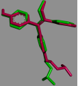
Redocking of the native 4-hydroxytamoxifen ligand into the ERα binding site of 3ERT reproduced the experimentally observed crystallographic pose with a root-mean-square deviation (RMSD) of 0.260 Å (Figure 1). This value is well below the generally accepted threshold of 2.0 Å for a reliable docking protocol , and the near-complete superimposition of the aromatic core, the alkyl side chain, and the dimethylaminoethoxy tail of the redocked and native poses confirms that the docking parameters faithfully reproduce the true binding geometry of the ERα ligand-binding pocket. This validated protocol was subsequently applied without modification to score the eight test phytochemicals and the reference drug, tamoxifen.
Figure 1. Superimposition of the native (pink) and redocked (green) pose of 4-hydroxytamoxifen within the ERα binding site (PDB: 3ERT). RMSD = 0.260 Å.
3.2. Molecular Docking of Test Compounds
All eight phytochemicals from Taraxacum officinale root docked successfully within the ERα ligand-binding pocket defined by the validated grid box. Binding scores ranged from -4.5 kcal/mol (lupeol) to -8.5 kcal/mol (luteolin), compared with -9.8 kcal/mol for tamoxifen, the reference drug (Table 1). Five compounds, luteolin (-8.5 kcal/mol), chicoric acid (-8.4 kcal/mol), quercetin (-8.0 kcal/mol), β-sitosterol (-7.9 kcal/mol), and chlorogenic acid (-7.4 kcal/mol), showed the most favourable docking energies among the test compounds, approaching but not matching the docking energy of tamoxifen. Luteolin was the strongest binder among the flavonoids, chicoric acid the strongest among the phenolic acids, and β-sitosterol the strongest among the triterpenoid/phytosterol subgroup. Taraxasterol (-5.6 kcal/mol), lupeol (-4.5 kcal/mol), and caffeic acid (-6.4 kcal/mol) showed less favourable docking energies and were not carried forward for detailed interaction analysis.
Table 1. Predicted binding scores of Taraxacum officinale phytochemicals and tamoxifen against ERα (PDB: 3ERT).

Compound

PubChem CID

Binding score (kcal/mol)

Taraxasterol (CPD1)

115250

-5.6

Lupeol (CPD2)

259846

-4.5

β-Sitosterol (CPD3)

222284

-7.9

Chlorogenic acid (CPD4)

1794427

-7.4

Caffeic acid (CPD5)

689043

-6.4

Chicoric acid (CPD6)

5281764

-8.4

Luteolin (CPD7)

5280445

-8.5

Quercetin (CPD8)

5280343

-8.0

Tamoxifen (reference drug)

2733526

-9.8

3.3. Protein-Ligand Interaction Analysis
Detailed two- and three-dimensional interaction analysis was performed for the five most promising phytochemicals (β-sitosterol, chlorogenic acid, chicoric acid, luteolin, and quercetin) and for tamoxifen, in both its PubChem-derived reference-drug docking and its native-ligand (4-hydroxytamoxifen) revalidation docking. A summary of the interaction profiles is presented in Table 2, followed by compound-specific descriptions and figures.
Table 2. Summary of key protein-ligand interactions within the ERα binding pocket.

Compound

H-bond residues

Hydrophobic/alkyl/pi contacts

Other notable interactions

β-Sitosterol (CPD3)

None observed

Leu346, Leu525, Ala350, Phe404, Leu387, Leu391, Leu384, Met388, Trp383, Met343

Purely hydrophobic binding mode; no polar anchoring

Chlorogenic acid (CPD4)

Glu353, Arg394

Ala350 (Pi-alkyl)

Two conventional H-bonds stabilise the caffeoyl carbonyl and quinic acid moiety

Chicoric acid (CPD6)

Gly521, His524, Glu419, Arg394

Leu387 (Pi-sigma), Leu525

Pi-sulfur contacts with Met421, Met343; most extensive H-bond network of all test compounds

Luteolin (CPD7)

Glu353, Glu419, His524, Gly420

Leu387, Leu384, Ala350 (Pi-alkyl)

Pi-sulfur with Met421

Quercetin (CPD8)

Glu353, Glu419, His524, Gly420

Leu387, Leu384, Ala350 (Pi-alkyl)

Pi-sulfur with Met421; additional B-ring hydroxyl vs. luteolin

Tamoxifen (PubChem reference)

None (ionic/aromatic anchoring)

Met421, Phe404, Leu346, Leu387, Ala350 (alkyl/Pi-alkyl)

Salt bridge and Pi-cation with Asp351 via protonated dimethylaminoethyl side chain

4-Hydroxytamoxifen (native, revalidation)

None (unfavourable contact noted)

Met421, Leu346, Leu387, Ala350, Leu349, Leu391 (alkyl/Pi-alkyl)

Unfavourable acceptor-acceptor clash with Glu353

β-Sitosterol (CPD3)
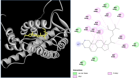
β-Sitosterol (-7.9 kcal/mol) occupied the ERα binding pocket entirely through hydrophobic contacts, engaging Leu346, Leu525, Ala350, Phe404, Leu387, Leu391, Leu384, Met388, Trp383, and Met343 via alkyl and Pi-alkyl interactions (Figure 2). No conventional hydrogen bonds were observed, indicating that the steroidal scaffold of β-sitosterol is accommodated within the hydrophobic sub-pocket of the receptor in a manner reminiscent of the non-polar core of tamoxifen's triphenylethylene skeleton, but without the polar anchoring contributed by tamoxifen's dimethylaminoethoxy side chain.
Figure 2. Predicted binding pose and 2D interaction diagram of β-sitosterol within the ERα binding pocket.
Chlorogenic acid (CPD4)
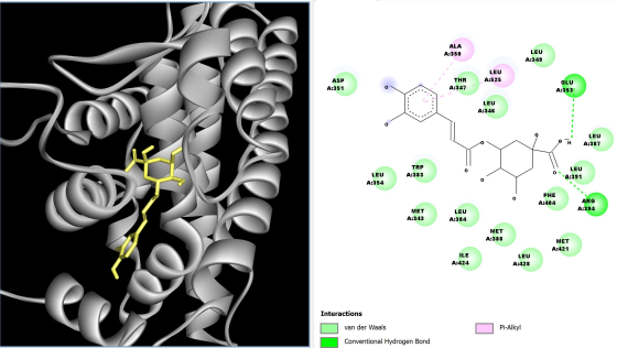
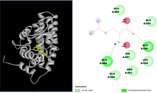
Chlorogenic acid (-7.4 kcal/mol) formed two conventional hydrogen bonds with Glu353 and Arg394, in addition to a Pi-alkyl contact with Ala350 (Figure 3). The caffeoyl-quinic acid ester structure of chlorogenic acid allowed its carbonyl and hydroxyl groups to engage in polar interactions with the receptor, a binding mode distinct from the purely hydrophobic engagement seen with β-sitosterol.
Figure 3. Predicted binding pose and 2D interaction diagram of chlorogenic acid within the ERα binding pocket.
Chicoric Acid (CPD6)
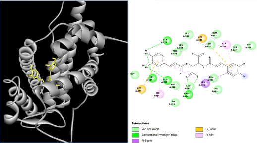
Chicoric acid (-8.4 kcal/mol), a dicaffeoyltartaric acid derivative, formed the most extensive polar interaction network among all test compounds, with conventional hydrogen bonds to Gly521, His524, Glu419, and Arg394, alongside Pi-sulfur interactions with Met421 and Met343 and a Pi-sigma interaction with Leu387 (Figure 4). The presence of two caffeoyl moieties in the chicoric acid structure appears to allow simultaneous polar and hydrophobic engagement with residues at both ends of the ERα binding pocket.
Figure 4. Predicted binding pose and 2D interaction diagram of chicoric acid within the ERα binding pocket.
Luteolin (CPD7)
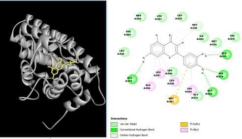
Luteolin (-8.5 kcal/mol), the strongest-binding phytochemical overall, formed conventional hydrogen bonds with Glu353, Glu419, His524, and Gly420, together with Pi-alkyl contacts (Leu387, Leu384, Ala350) and a Pi-sulfur interaction with Met421 (Figure 5). The flavone backbone of luteolin, bearing hydroxyl substituents at the 5-, 7-, 3′-, and 4′-positions, positioned its catechol-like B-ring hydroxyls to donate hydrogen bonds to Glu419 and His524, closely paralleling the hydrogen-bonding pattern seen for the native 4-hydroxytamoxifen ligand at the same receptor site.
Figure 5. Predicted binding pose and 2D interaction diagram of luteolin within the ERα binding pocket.
Quercetin (CPD8)
Quercetin (-8.0 kcal/mol) shares an identical flavone core with luteolin, differing only by an additional hydroxyl group on the B-ring, and accordingly produced a near-identical interaction fingerprint: conventional hydrogen bonds with Glu353, Glu419, His524, and Gly420, Pi-alkyl contacts with Leu387, Leu384, and Ala350, and a Pi-sulfur interaction with Met421 (Figure 6). The additional hydroxyl substituent did not markedly change the binding score relative to luteolin, but the conserved hydrogen-bonding network across both flavonoids reinforces the reproducibility of this polar anchoring mode within the ERα pocket.
Figure 6. Predicted binding pose and 2D interaction diagram of quercetin within the ERα binding pocket, including a Pi-sulfur interaction with Met421.
Tamoxifen (Reference Drug, PubChem)
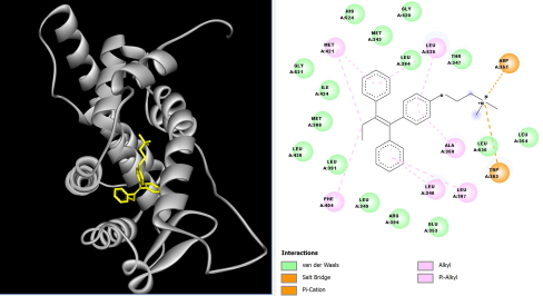
Tamoxifen (-9.8 kcal/mol), docked as the reference drug using its PubChem-derived structure, engaged the ERα pocket through an extensive alkyl and Pi-alkyl network (Met421, Phe404, Leu346, Leu387, Ala350) together with a salt bridge and Pi-cation interaction between its protonated dimethylaminoethyl side chain and Asp351 (Figure 7). This ionic anchoring interaction, absent from all eight phytochemicals evaluated, is consistent with the established mechanism by which SERM side chains engage Asp351 to displace helix 12 and antagonise receptor activation .
Figure 7. Predicted binding pose and 2D interaction diagram of tamoxifen (PubChem CID 2733526) within the ERα binding pocket, showing salt bridge/Pi-cation interaction with Asp351.
4-Hydroxytamoxifen (Native Ligand, Revalidation Docking)
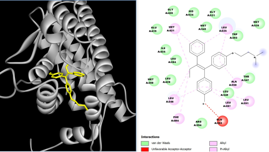
As part of the docking validation exercise, the native ligand extracted from 3ERT (4-hydroxytamoxifen) was independently redocked and its interaction profile examined. This pose reproduced the same alkyl/Pi-alkyl hydrophobic network observed for the PubChem tamoxifen docking (Met421, Leu346, Leu387, Ala350, Leu349, Leu391) but showed an unfavourable acceptor-acceptor clash with Glu353 involving the ligand's phenolic hydroxyl group (Figure 8). This finding is consistent with known steric sensitivity of the 4-hydroxy substituent within this sub-pocket and does not detract from the high fidelity of the redocking RMSD, since RMSD reflects positional accuracy rather than interaction favourability.
Figure 8. Predicted binding pose and 2D interaction diagram of the redocked native ligand (4-hydroxytamoxifen) within the ERα binding pocket, showing an unfavourable acceptor-acceptor interaction with Glu353.
3.4. Drug-Likeness, ADMET, and Toxicity Profile
All eight phytochemicals and tamoxifen were evaluated for physicochemical drug-likeness, pharmacokinetic behaviour, and predicted toxicity (Tables 3 and 4). Six of the eight test compounds carried zero or one Lipinski Rule-of-Five violations, indicating broadly favourable oral drug-likeness across the panel; taraxasterol, lupeol, and β-sitosterol each carried one Lipinski violation on account of elevated LogP. Chlorogenic acid, caffeic acid, luteolin, and quercetin recorded zero violations, matching or exceeding the drug-likeness profile of tamoxifen itself, which carried one violation (LogP = 6.06).
Predicted human intestinal absorption (HIA) varied considerably across the panel. The triterpenoid/phytosterol compounds (taraxasterol, lupeol, and β-sitosterol) showed high predicted HIA (94.25-94.80%), comparable to tamoxifen (96.89%), consistent with their high lipophilicity (LogP 5.82-7.91). Caffeic acid (69.40%) and quercetin (75.20%) showed moderate predicted absorption, luteolin showed similarly moderate absorption (81.13%), while chlorogenic acid (36.30%) and, most markedly, chicoric acid (0.69%) showed poor predicted intestinal absorption, reflecting their high topological polar surface area (164.75 Å2 and 208.12 Å2, respectively) and multiple ionisable carboxylic acid groups. None of the nine compounds, including tamoxifen, were predicted to cross the blood-brain barrier, which is a favourable safety feature for compounds intended to act peripherally on breast tissue ERα rather than centrally.
Cytochrome P450 interaction predictions distinguished the flavonoids from the remaining phytochemicals: luteolin was predicted to inhibit CYP1A2 and CYP2C9, and quercetin was predicted to inhibit CYP1A2, CYP2C9, and CYP2C19, raising a theoretical potential for drug-drug interactions upon co-administration with substrates of these isoforms. Tamoxifen itself was predicted to inhibit CYP1A2 and CYP3A4, the latter being the isoform principally responsible for its own metabolic activation to endoxifen , whereas none of the triterpenoid, phytosterol, or simple phenolic acid compounds (taraxasterol, lupeol, β-sitosterol, chlorogenic acid, caffeic acid, chicoric acid) showed predicted inhibition of any of the five CYP isoforms evaluated.
Acute oral toxicity predictions indicated favourable safety margins for most test compounds. Taraxasterol, chlorogenic acid, and chicoric acid showed the highest predicted LD50 values (5000 mg/kg, GHS Class 5, the least hazardous classification), followed by caffeic acid (2980 mg/kg, Class 5) and lupeol (2000 mg/kg, Class 4). β-Sitosterol (890 mg/kg, Class 4) and luteolin (3919 mg/kg, Class 5) also fell within acceptable ranges. Quercetin showed the lowest predicted LD50 among the phytochemicals (159 mg/kg, GHS Class 3), placing it in a more hazardous acute toxicity category than the other seven compounds, though still less severe than the most acutely toxic GHS classes. Tamoxifen showed a predicted LD50 of 1190 mg/kg (GHS Class 4) and was the only compound in the panel flagged as having an active hepatotoxicity liability; all eight phytochemicals were predicted to be hepatotoxicity-inactive.
Table 3. Physicochemical properties and drug-likeness parameters (SwissADME).

Ligand

MW (g/mol)

LogP

TPSA (Å2)

Lipinski viol.

HIA (%)

Caco-2 (log Papp)

BBB (log BB)

Taraxasterol

426.72

5.82

20.23

1

94.25

1.21

No

Lupeol

426.72

7.91

20.23

1

94.80

1.23

No

β-Sitosterol

414.71

7.78

20.23

1

94.50

1.20

No

Chlorogenic acid

354.31

-0.71

164.75

0

36.30

-0.84

No

Caffeic acid

180.16

0.92

77.76

0

69.40

0.60

No

Chicoric acid

474.37

1.01

208.12

1

0.69

-1.12

No

Luteolin

286.24

1.10

111.13

0

81.13

0.10

No

Quercetin

302.24

0.79

131.36

0

75.20

0.18

No

Tamoxifen

371.51

6.06

12.47

1

96.89

1.10

No

Table 4. Predicted pharmacokinetic, cytochrome P450 interaction, and toxicity parameters (pkCSM).

Ligand

Vss (log L/kg)

CYP1A2

CYP2C9

CYP2C19

CYP2D6

CYP3A4

Cl. (log mL/min/kg)

LD50 (mg/kg)

GHS class

Hepatox.

Taraxasterol

0.13

No

No

No

No

No

0.12

5000

5

Inactive

Lupeol

0.12

No

No

No

No

No

0.15

2000

4

Inactive

β-Sitosterol

0.20

No

No

No

No

No

0.63

890

4

Inactive

Chlorogenic acid

-0.58

No

No

No

No

No

0.30

5000

5

Inactive

Caffeic acid

-1.0

No

No

No

No

No

0.51

2980

5

Inactive

Chicoric acid

0.09

No

No

No

No

No

0.08

5000

5

Inactive

Luteolin

0.12

Yes

Yes

No

No

No

0.5

3919

5

Inactive

Quercetin

-0.23

Yes

Yes

Yes

No

No

0.42

159

3

Inactive

Tamoxifen

0.83

Yes

No

No

No

Yes

0.60

1190

4

Active

4. Discussion
The present study set out to determine whether individual, chemically defined phytochemicals from Taraxacum officinale root show molecular interactions with estrogen receptor alpha, providing a receptor-level complement to previous extract-based reports of dandelion's anticancer activity. The redocking validation, which reproduced the crystallographic pose of 4-hydroxytamoxifen with an RMSD of 0.260 Å, provides a high degree of confidence that the docking energies and interaction profiles reported here reflect geometrically reliable predictions of ligand pose within the ERα binding pocket, rather than artefacts of an unvalidated docking protocol .
Among the eight phytochemicals evaluated, luteolin, chicoric acid, quercetin, β-sitosterol, and chlorogenic acid emerged as the compounds with the most favourable predicted ERα docking energies (-7.4 to -8.5 kcal/mol), approaching the docking energy calculated for tamoxifen (-9.8 kcal/mol). Notably, the two flavonoids, luteolin and quercetin, reproduced a hydrogen-bonding pattern similar to that of the native 4-hydroxytamoxifen ligand, engaging Glu419 and His524 through their catechol-like B-ring hydroxyls. This convergence in predicted binding mode suggests that the flavone scaffold, independent of the steroidal or triphenylethylene chemotypes typical of estrogenic and anti-estrogenic ligands, may be capable of accessing the same polar recognition elements of the ERα ligand-binding domain that underlie tamoxifen's antagonist activity, although docking energies and shared contact residues indicate potential interaction rather than demonstrated biological efficacy. This observation is consistent with a substantial body of prior work establishing flavonoids such as quercetin and luteolin as phytoestrogen-like ERα ligands capable of both agonist and antagonist behaviour depending on cellular and structural context .
Chicoric acid's comparatively favourable docking energy (-8.4 kcal/mol) and extensive hydrogen-bonding network (the most polar-interaction-rich profile among all eight phytochemicals) is consistent with its dicaffeoyltartaric acid structure, which presents two catechol-like caffeoyl termini capable of simultaneous polar engagement at opposite ends of the binding pocket. This is the same class of compound previously highlighted in reviews of Taraxacum-derived oncotherapy candidates as a promising tumour-modulating constituent , and the present docking data provide, to our knowledge, the first structural evidence suggesting that chicoric acid may interact with the ERα ligand-binding domain specifically, rather than acting solely through the non-receptor-mediated apoptotic or oxidative-stress mechanisms previously reported for crude dandelion extracts ; this predicted interaction would need to be confirmed experimentally.
β-Sitosterol's binding mode, by contrast, was purely hydrophobic, lacking any hydrogen-bond anchoring within the pocket. This is consistent with its steroidal, non-polar structure, which lacks the phenolic or carboxylic acid groups available to the flavonoids and phenolic acids in this panel. While its binding score (-7.9 kcal/mol) was competitive, the absence of polar anchoring residues typically associated with high-affinity, selective ERα antagonism raises the possibility that its predicted affinity reflects a more generic hydrophobic-pocket fit rather than the specific polar recognition pattern that distinguishes established SERMs. This distinction underscores the value of interaction-level analysis over binding score alone when prioritising candidates for further study.
Taraxasterol, lupeol, and caffeic acid showed the least favourable docking energies in the panel (-5.6, -4.5, and -6.4 kcal/mol, respectively) and were therefore not carried forward to detailed interaction analysis. These differences are plausibly explained by molecular structure. Taraxasterol and lupeol are both large, rigid pentacyclic triterpenoids that, unlike β-sitosterol, lack a flexible side chain capable of extending toward polar residues at the periphery of the ERα pocket; their bulk may also restrict optimal orientation within the binding cavity, limiting the number of favourable contacts relative to their molecular size. Caffeic acid, by contrast, is the smallest phenolic acid in the panel (180.16 g/mol) and possesses only a single catechol ring with no ester linkage to a second polar moiety, unlike chlorogenic acid (which adds a quinic acid ester) or chicoric acid (which presents two caffeoyl termini); this more limited polar surface area and reduced molecular complexity likely restrict caffeic acid to fewer simultaneous contacts within the pocket. Their comparatively modest predicted affinity for ERα does not exclude a role in dandelion's previously reported anticancer activity, since non-ERα mechanisms (including direct pro-apoptotic, pro-autophagic, and oxidative-stress-mediated pathways) have been implicated in the cytotoxic effects of dandelion root extract in prior cell-based studies . Rather, these findings suggest that if dandelion root extract exerts any ERα-mediated component of anticancer activity, this is more plausibly attributable to its flavonoid and phenolic acid constituents than to its triterpenoid fraction.
Critically, no phytochemical in this panel reproduced tamoxifen's ionic anchoring interaction with Asp351, mediated by the protonated dimethylaminoethyl side chain that is characteristic of triphenylethylene SERMs . This structural feature is widely regarded as central to the specific, high-affinity antagonism exhibited by tamoxifen and related SERMs, since it positions the ligand precisely to displace helix 12 and block coactivator recruitment. Its absence across all eight test compounds indicates that, while several dandelion phytochemicals, particularly the flavonoids and chicoric acid, show predicted molecular interactions consistent with ERα engagement, none is likely to reproduce tamoxifen's mechanism of action with equivalent specificity. These docking results indicate potential interaction with the ERα ligand-binding domain that warrants experimental validation, rather than demonstrating that these compounds function as ERα antagonists or possess anticancer efficacy.
From a translational perspective, the ADMET and toxicity profile of the panel is broadly encouraging in silico. Chlorogenic acid, caffeic acid, luteolin, and quercetin each showed zero Lipinski violations, a physicochemical drug-likeness profile at least as favourable as that of tamoxifen . None of the nine compounds evaluated, including tamoxifen, was predicted to cross the blood-brain barrier, a favourable feature for a receptor target expressed principally in peripheral hormone-responsive tissue. The predicted absence of hepatotoxicity across all eight phytochemicals, contrasting with tamoxifen's own flagged hepatotoxicity liability, is a notable finding given that tamoxifen-associated hepatotoxicity, including rare cases of hepatocellular injury and an increased risk of non-alcoholic fatty liver changes, is a recognised, if uncommon, clinical concern . These compounds therefore displayed more favourable predicted hepatotoxicity profiles in silico than tamoxifen; whether this translates into an improved hepatic safety margin in vivo would need to be confirmed experimentally, since in silico hepatotoxicity models do not capture dose, exposure duration, or metabolic activation in a living system.
The most important caveat concerns quercetin's predicted acute oral toxicity (LD50 = 159 mg/kg, GHS Class 3), which places it in a more hazardous category than every other phytochemical evaluated, despite its otherwise favourable drug-likeness and ERα-binding profile. Quercetin and luteolin were also the only two phytochemicals flagged for potential CYP1A2 and CYP2C9 inhibition (with quercetin additionally flagged for CYP2C19 inhibition), raising a theoretical risk of pharmacokinetic drug-drug interactions. These findings do not disqualify quercetin as a compound of interest, since in silico LD50 predictions carry considerable uncertainty and quercetin has an extensive history of dietary and supplemental human exposure at far lower doses than those associated with acute toxicity thresholds in rodent models, but they do indicate that any future experimental work with quercetin in this context should include a careful dose-ranging toxicological assessment.
Taken together, these findings help to narrow the mechanistic space underlying dandelion root's reported anticancer activity. Prior cell-based studies established extract-level cytotoxicity in colorectal and pancreatic cancer cell lines but did not identify a specific molecular target. The present in silico analysis provides structural evidence that at least five individual T. officinale phytochemicals (β-sitosterol, chlorogenic acid, chicoric acid, luteolin, and quercetin) show docking energies and, in several cases, hydrogen-bonding patterns comparable to a validated SERM reference standard, suggesting potential interaction with the ERα ligand-binding domain that warrants experimental validation. Rather than dandelion root acting as an undifferentiated cytotoxic agent, these data point to specific constituents, particularly the flavonoids, as candidates for targeted experimental follow-up, rather than the extract as a whole.
5. Conclusions
This study employed a validated molecular docking protocol (redocking RMSD = 0.260 Å) to evaluate eight bioactive phytochemicals from Taraxacum officinale root as potential estrogen receptor alpha inhibitors, benchmarked against tamoxifen. Luteolin, chicoric acid, quercetin, β-sitosterol, and chlorogenic acid showed the most favourable docking energies, with luteolin and quercetin in particular reproducing key hydrogen-bonding interactions characteristic of the native ERα ligand within the same pocket. None of the eight phytochemicals reproduced tamoxifen's signature ionic interaction with Asp351, indicating that these compounds are unlikely to act as direct tamoxifen mimetics, but several nonetheless show docking patterns consistent with potential ERα interaction that warrants experimental validation. Drug-likeness and ADMET profiling further identified chlorogenic acid, caffeic acid, luteolin, and quercetin as possessing favourable oral drug-likeness with zero Lipinski violations, and all eight phytochemicals displayed more favourable predicted hepatotoxicity profiles in silico than tamoxifen. Quercetin's comparatively lower predicted LD50 warrants caution and further scrutiny in any future experimental work. Collectively, these findings offer a receptor-level perspective on dandelion root's reported anticancer activity and identify specific phytochemical candidates, most notably luteolin, quercetin, and chicoric acid, for prioritised experimental validation, including in vitro ERα binding assays, transcriptional reporter assays, and cell-based proliferation studies in ER-positive breast cancer models, before any conclusions regarding Taraxacum officinale's therapeutic relevance to hormone receptor-positive breast cancer can be drawn.
Abbreviations

ERα

Estrogen Receptor Alpha

SERM

Selective Estrogen Receptor Modulator

PDB

Protein Data Bank

RMSD

Root-Mean-Square Deviation

ADMET

Absorption, Distribution, Metabolism, Excretion, and Toxicity

TPSA

Topological Polar Surface Area

HIA

Human Intestinal Absorption

BBB

Blood-Brain Barrier

CYP

Cytochrome P450

LD50

Median Lethal Dose

GHS

Globally Harmonised System

MW

Molecular Weight

CID

PubChem Compound Identifier

SDF

Structure-Data File

PDBQT

Protein Data Bank, Partial Charge (Q), and Atom Type (T) File Format

Vss

Steady-State Volume of Distribution

Papp

Apparent Permeability

TNBC

Triple-Negative Breast Cancer

HER2

Human Epidermal Growth Factor Receptor 2

ER

Estrogen Receptor

PR

Progesterone Receptor

ESR1

Estrogen Receptor 1 (gene)

AF

Activation Function

GLOBOCAN

Global Cancer Observatory

Acknowledgments
I sincerely acknowledge Mrs Modupe Ajike Lawrence and Mr. Ayodele Idris Aminu for the support and encouragement received during the course of this work.
Author Contributions
Osakuade Ayodeji Sunday: Conceptualization, Data curation, Formal Analysis, Investigation, Methodology, Software, Visualization, Writing – original draft, Writing – review & editing
Ikor Kelvin Okibe: Methodology, Supervision, Validation, Writing – review & editing
Data Availability Statement
The data supporting the outcome of this research work has been reported in this manuscript.
Conflicts of Interest
The authors declare no conflicts of interest.
References
[1] Bray, F., Laversanne, M., Sung, H., Ferlay, J., Siegel, R. L., Soerjomataram, I., Jemal, A. Global Cancer Statistics 2022: GLOBOCAN Estimates of Incidence and Mortality Worldwide for 36 Cancers in 185 Countries. CA: A Cancer Journal for Clinicians. 2024, 74(3), 229-263.
[2] Freihat, O., Sipos, D., Kovacs, A. Global Burden and Projections of Breast Cancer Incidence and Mortality to 2050: A Comprehensive Analysis of GLOBOCAN Data. Frontiers in Public Health. 2025, 13, 1622954.
[3] Zagami, P., Carey, L. A. Triple Negative Breast Cancer: Pitfalls and Progress. NPJ Breast Cancer. 2022, 8(1), 95.
[4] Fernando, T. M., Moore, H. M., Wongchenko, M. J., Metcalfe, C. Next-Generation Estrogen Receptor-Targeted Therapeutics. Annual Review of Cancer Biology. 2023, 7, 313-330.
[5] Chen, P., Li, B., Ou-Yang, L. Role of Estrogen Receptors in Health and Disease. Frontiers in Endocrinology. 2022, 13, 839005.
[6] Shiau, A. K., Barstad, D., Loria, P. M., Cheng, L., Kushner, P. J., Agard, D. A., Greene, G. L. The Structural Basis of Estrogen Receptor/Coactivator Recognition and the Antagonism of This Interaction by Tamoxifen. Cell. 1998, 95(7), 927-937.
[7] Will, M., Liang, J., Metcalfe, C., Chandarlapaty, S. Therapeutic Resistance to Anti-Oestrogen Therapy in Breast Cancer. Nature Reviews Cancer. 2023, 23(10), 673-685.
[8] Ozyurt, R., Ozpolat, B. Molecular Mechanisms of Anti-Estrogen Therapy Resistance and Novel Targeted Therapies. Cancers. 2022, 14(21), 5206.
[9] Yao, J., Deng, K., Huang, J., Zeng, R., Zuo, J. Progress in the Understanding of the Mechanism of Tamoxifen Resistance in Breast Cancer. Frontiers in Pharmacology. 2020, 11, 592912.
[10] Ghanavati, M., Khorshidi, Y., Shadnoush, M., Akbari, M. E., Ardehali, S. H., Chavarri-Guerra, Y., Akbari, A., Barragan-Carrillo, R., Amin Amlashi, M., Javid, Z., Rahmani, J. Tamoxifen Use and Risk of Endometrial Cancer in Breast Cancer Patients: A Systematic Review and Dose-Response Meta-Analysis. Cancer Reports. 2023, 6(6), e1806.
[11] Jia, J., Tian, C., Han, W., Ma, Q. Real-World Assessment of Thromboembolic Risk Associated with Tamoxifen. Scientific Reports. 2025, 15, 27820.
[12] Asma, S. T., Acaroz, U., Imre, K., Morar, A., Shah, S. R. A., Hussain, S. Z., Arslan-Acaroz, D., Demirbas, H., Hajrulai-Musliu, Z., Istanbullugil, F. R., Soleimanzadeh, A., Morozov, D., Zhu, K., Herman, V., Ayad, A., Athanassiou, C., Ince, S. Natural Products/Bioactive Compounds as a Source of Anticancer Drugs. Cancers. 2022, 14(24), 6203.
[13] Chavda, V. P., Ertas, Y. N., Walhekar, V., Modh, D., Doshi, A., Shah, N., Anand, K., Chhabria, M. Advanced Computational Methodologies Used in the Discovery of New Natural Anticancer Compounds. Frontiers in Pharmacology. 2021, 12, 702611.
[14] Wu, J., Sun, J., Liu, M., Zhang, X., Kong, L., Ma, L., Jiang, S., Liu, X., Ma, W. Botany, Traditional Use, Phytochemistry, Pharmacology and Quality Control of Taraxaci Herba: Comprehensive Review. Pharmaceuticals. 2024, 17(9), 1113.
[15] Al-Eisawi, Z., Abderrahman, S. M., Al-khalaf, I. F., Al-Abbassi, R., Bustanji, Y. K. Taraxacum officinale Extracts Exhibit Safe and Selective Anticancer Activity. Natural Product Journal. 2022, 12(1), 69-77.
[16] Tanasa Acretei, M. V., Negreanu-Pirjol, T., Olariu, L., Negreanu-Pirjol, B. S., Lepadatu, A. C., Anghel Cireasa, L. Bioactive Compounds from Vegetal Organs of Taraxacum Species (Dandelion) with Biomedical Applications: A Review. International Journal of Molecular Sciences. 2025, 26(2), 450.
[17] Ovadje, P., Ammar, S., Guerrero, J. A., Arnason, J. T., Pandey, S. Dandelion Root Extract Affects Colorectal Cancer Proliferation and Survival Through the Activation of Multiple Death Signalling Pathways. Oncotarget. 2016, 7(45), 73080-73100.
[18] Ovadje, P., Chochkeh, M., Akbari-Asl, P., Hamm, C., Pandey, S. Selective Induction of Apoptosis and Autophagy Through Treatment with Dandelion Root Extract in Human Pancreatic Cancer Cells. Pancreas. 2012, 41(7), 1039-1047.
[19] Bugnon, M., Röhrig, U. F., Goullieux, M., Perez, M. A. S., Daina, A., Michielin, O., Zoete, V. SwissDock 2024: Major Enhancements for Small-Molecule Docking with Attracting Cavities and AutoDock Vina. Nucleic Acids Research. 2024, 52(W1), W324-W332.
[20] Agu, P. C., Afiukwa, C. A., Orji, O. U., Ezeh, E. M., Ofoke, I. H., Ogbu, C. O., Ugwuja, E. I., Aja, P. M. Molecular Docking as a Tool for the Discovery of Molecular Targets of Nutraceuticals in Diseases Management. Scientific Reports. 2023, 13(1), 13398.
[21] Lipinski, C. A., Lombardo, F., Dominy, B. W., Feeney, P. J. Experimental and Computational Approaches to Estimate Solubility and Permeability in Drug Discovery and Development Settings. Advanced Drug Delivery Reviews. 2001, 46(1-3), 3-26.
[22] Daina, A., Michielin, O., Zoete, V. SwissADME: A Free Web Tool to Evaluate Pharmacokinetics, Drug-Likeness and Medicinal Chemistry Friendliness of Small Molecules. Scientific Reports. 2017, 7, 42717.
[23] Pires, D. E. V., Blundell, T. L., Ascher, D. B. pkCSM: Predicting Small-Molecule Pharmacokinetic and Toxicity Properties Using Graph-Based Signatures. Journal of Medicinal Chemistry. 2015, 58(9), 4066-4072.
[24] Burley, S. K., Bhikadiya, C., Bi, C., Bittrich, S., Chen, L., Crichlow, G. V., Christie, C. H., Dalenberg, K., Di Costanzo, L., Duarte, J. M., Dutta, S., Feng, Z., Ganesan, S., Goodsell, D. S., Ghosh, S., Green, R. K., Guranović, V., Guzenko, D., Hudson, B. P., Lawson, C. L., Liang, Y., Lowe, R., Namkoong, H., Peisach, E., Persikova, I., Randle, C., Rose, A., Rose, Y., Sali, A., Segura, J., Sekharan, M., Shao, C., Tao, Y. P., Voigt, M., Westbrook, J. D., Young, J. Y., Zardecki, C., Zhuravleva, M. RCSB Protein Data Bank: Powerful New Tools for Exploring 3D Structures of Biological Macromolecules for Basic and Applied Research and Education in Fundamental Biology, Biomedicine, Biotechnology, Bioengineering and Energy Sciences. Nucleic Acids Research. 2021, 49(D1), D437-D451.
[25] Kim, S., Chen, J., Cheng, T., Gindulyte, A., He, J., He, S., Li, Q., Shoemaker, B. A., Thiessen, P. A., Yu, B., Zaslavsky, L., Zhang, J., Bolton, E. E. PubChem in 2021: New Data Content and Improved Web Interfaces. Nucleic Acids Research. 2021, 49(D1), D1388-D1395.
[26] DeLano, W. L. The PyMOL Molecular Graphics System. Schrödinger, LLC. 2002.
[27] Morris, G. M., Huey, R., Lindstrom, W., Sanner, M. F., Belew, R. K., Goodsell, D. S., Olson, A. J. AutoDock4 and AutoDockTools4: Automated Docking with Selective Receptor Flexibility. Journal of Computational Chemistry. 2009, 30(16), 2785-2791.
[28] Eberhardt, J., Santos-Martins, D., Tillack, A. F., Forli, S. AutoDock Vina 1.2.0: New Docking Methods, Expanded Force Field, and Python Bindings. Journal of Chemical Information and Modeling. 2021, 61(8), 3891-3898.
[29] Sanchez-Spitman, A. B., Swen, J. J., Dezentje, V. O., Moes, D., Gelderblom, H., Guchelaar, H. J. Clinical Pharmacokinetics and Pharmacogenetics of Tamoxifen and Endoxifen. Expert Review of Clinical Pharmacology. 2019, 12(6), 523-536.
[30] Cord, D., Rîmbu, M. C., Popescu, L. New Prospects in Oncotherapy: Bioactive Compounds from Taraxacum officinale. Medicine and Pharmacy Reports. 2025, 98(3), 290-299.
[31] Osborne, C. K. Tamoxifen in the Treatment of Breast Cancer. New England Journal of Medicine. 1998, 339(22), 1609-1618.
Cite This Article
  • APA Style

    Sunday, O. A., Okibe, I. K. (2026). Molecular Docking, Drug-Likeness, and Toxicity Profiling of Taraxacum Officinale-derived Phytochemicals Against Estrogen Receptor Alpha (ERα) in Breast Cancer. Computational Biology and Bioinformatics, 14(2), 54-69. https://doi.org/10.11648/j.cbb.20261402.11

    Copy | Download

    ACS Style

    Sunday, O. A.; Okibe, I. K. Molecular Docking, Drug-Likeness, and Toxicity Profiling of Taraxacum Officinale-derived Phytochemicals Against Estrogen Receptor Alpha (ERα) in Breast Cancer. Comput. Biol. Bioinform. 2026, 14(2), 54-69. doi: 10.11648/j.cbb.20261402.11

    Copy | Download

    AMA Style

    Sunday OA, Okibe IK. Molecular Docking, Drug-Likeness, and Toxicity Profiling of Taraxacum Officinale-derived Phytochemicals Against Estrogen Receptor Alpha (ERα) in Breast Cancer. Comput Biol Bioinform. 2026;14(2):54-69. doi: 10.11648/j.cbb.20261402.11

    Copy | Download

  • @article{10.11648/j.cbb.20261402.11,
      author = {Osakuade Ayodeji Sunday and Ikor Kelvin Okibe},
      title = {Molecular Docking, Drug-Likeness, and Toxicity Profiling of Taraxacum Officinale-derived Phytochemicals Against Estrogen Receptor Alpha (ERα) in Breast Cancer},
      journal = {Computational Biology and Bioinformatics},
      volume = {14},
      number = {2},
      pages = {54-69},
      doi = {10.11648/j.cbb.20261402.11},
      url = {https://doi.org/10.11648/j.cbb.20261402.11},
      eprint = {https://article.sciencepublishinggroup.com/pdf/10.11648.j.cbb.20261402.11},
      abstract = {Public interest in the anticancer potential of dandelion (Taraxacum officinale) root has grown following reports that its extract can induce cell death in cancer cell lines, but whether individual T. officinale phytochemicals directly engage estrogen receptor alpha (ERα), the principal target in hormone receptor-positive breast cancer, has not been characterised. This study computationally evaluated eight bioactive phytochemicals reported from T. officinale root (taraxasterol, lupeol, β-sitosterol, chlorogenic acid, caffeic acid, chicoric acid, luteolin, and quercetin) as potential ERα inhibitors, benchmarked against tamoxifen. Molecular docking was performed using AutoDock Vina against the ERα ligand-binding domain (PDB: 3ERT), with docking protocol validity confirmed by redocking the native ligand and calculating the root-mean-square deviation (RMSD). Drug-likeness was assessed using SwissADME (Lipinski's Rule of Five), and pharmacokinetic and toxicity behaviour were predicted using pkCSM. Redocking reproduced the native ligand pose with an RMSD of 0.260 Å. Binding scores ranged from -4.5 (lupeol) to -8.5 kcal/mol (luteolin), versus -9.8 kcal/mol for tamoxifen. Luteolin, quercetin, chicoric acid, β-sitosterol, and chlorogenic acid showed the most favourable docking energies; luteolin and quercetin reproduced key hydrogen bonds seen for the native ligand, though none reproduced tamoxifen's Asp351 salt bridge. Chlorogenic acid, caffeic acid, luteolin, and quercetin showed zero Lipinski violations, and all eight phytochemicals were hepatotoxicity-inactive in silico, unlike tamoxifen. These findings offer a receptor-level perspective on dandelion's reported anticancer activity and identify candidates warranting experimental validation.},
     year = {2026}
    }
    

    Copy | Download

  • TY  - JOUR
    T1  - Molecular Docking, Drug-Likeness, and Toxicity Profiling of Taraxacum Officinale-derived Phytochemicals Against Estrogen Receptor Alpha (ERα) in Breast Cancer
    AU  - Osakuade Ayodeji Sunday
    AU  - Ikor Kelvin Okibe
    Y1  - 2026/10/09
    PY  - 2026
    N1  - https://doi.org/10.11648/j.cbb.20261402.11
    DO  - 10.11648/j.cbb.20261402.11
    T2  - Computational Biology and Bioinformatics
    JF  - Computational Biology and Bioinformatics
    JO  - Computational Biology and Bioinformatics
    SP  - 54
    EP  - 69
    PB  - Science Publishing Group
    SN  - 2330-8281
    UR  - https://doi.org/10.11648/j.cbb.20261402.11
    AB  - Public interest in the anticancer potential of dandelion (Taraxacum officinale) root has grown following reports that its extract can induce cell death in cancer cell lines, but whether individual T. officinale phytochemicals directly engage estrogen receptor alpha (ERα), the principal target in hormone receptor-positive breast cancer, has not been characterised. This study computationally evaluated eight bioactive phytochemicals reported from T. officinale root (taraxasterol, lupeol, β-sitosterol, chlorogenic acid, caffeic acid, chicoric acid, luteolin, and quercetin) as potential ERα inhibitors, benchmarked against tamoxifen. Molecular docking was performed using AutoDock Vina against the ERα ligand-binding domain (PDB: 3ERT), with docking protocol validity confirmed by redocking the native ligand and calculating the root-mean-square deviation (RMSD). Drug-likeness was assessed using SwissADME (Lipinski's Rule of Five), and pharmacokinetic and toxicity behaviour were predicted using pkCSM. Redocking reproduced the native ligand pose with an RMSD of 0.260 Å. Binding scores ranged from -4.5 (lupeol) to -8.5 kcal/mol (luteolin), versus -9.8 kcal/mol for tamoxifen. Luteolin, quercetin, chicoric acid, β-sitosterol, and chlorogenic acid showed the most favourable docking energies; luteolin and quercetin reproduced key hydrogen bonds seen for the native ligand, though none reproduced tamoxifen's Asp351 salt bridge. Chlorogenic acid, caffeic acid, luteolin, and quercetin showed zero Lipinski violations, and all eight phytochemicals were hepatotoxicity-inactive in silico, unlike tamoxifen. These findings offer a receptor-level perspective on dandelion's reported anticancer activity and identify candidates warranting experimental validation.
    VL  - 14
    IS  - 2
    ER  - 

    Copy | Download

Author Information
  • Department of Biotechnology, Nile University of Nigeria, Abuja, Nigeria

    Biography: Osakuade Ayodeji Sunday is a Biotechnology researcher with a background in Plant Science and Biotechnology. He holds a Bachelor of Science degree in Plant Science and Biotechnology and is currently pursuing a Master of Science degree in Biotechnology at Nile University of Nigeria. His research interests span computational drug discovery, bioinformatics, molecular modelling, molecular docking, molecular dynamics simulations, antimicrobial resistance, and the evaluation of natural products for therapeutic applications. He is particularly interested in applying computational and data driven approaches to investigate biological systems, identify potential therapeutic targets, and support early stage drug discovery. His broader research interests also include biotechnology, biomedical research, and the application of computational tools to address challenges in health and disease.

    Research Fields: Computational Drug Discovery, Bioinformatics, biotechnology, biomedical research, Molecular Modelling, Molecular Dynamics Simulation, Antimicrobial Resistance, Plant Biotechnology, Ethnomedicine, Natural Products Research, Breast Cancer Research, Biotechnology


  • Department of Healthcare& Research, Adentrix Vetrex Concepts, Abuja, Nigeria

    Biography: Ikor Kelvin Okibe is a Biotechnology researcher and MSc candidate at Nile University of Nigeria, Abuja, with a Bachelor of Science degree in Biology from Benue State University. He is a dedicated member of the Biotechnology Society of Nigeria (BSN) and is actively engaged in biotechnology research and its practical applications. His research interests span biomedical biotechnology, molecular biology, genomics, gene therapy, phytochemistry, and computational drug discovery. His current research focuses on the therapeutic potential of bioactive compounds from Vernonia amygdalina (bitter leaf) for the management of type 2 diabetes, integrating in silico molecular docking and in vivo approaches. His broader interests include phytochemical research, molecular docking, enzymatic inhibition, metabolic disorders, genomics, gene therapy, and the development of innovative biotechnology-based therapeutic approaches.

    Research Fields: Biomedical Biotechnology, Molecular Biology, Genomics, Gene Therapy, Computational Drug Discovery, Phytochemistry, Molecular Docking, Plant Biotechnology, Diabetes Research

  • Abstract
  • Keywords
  • Document Sections

    1. 1. Introduction
    2. 2. Materials and Methods
    3. 3. Results
    4. 4. Discussion
    5. 5. Conclusions
    Show Full Outline
  • Abbreviations
  • Acknowledgments
  • Author Contributions
  • Data Availability Statement
  • Conflicts of Interest
  • References
  • Cite This Article
  • Author Information